Figure 1.
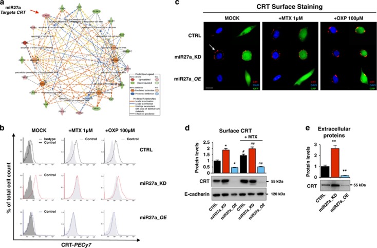
Calreticulin cell surface exposure is downregulated by miR-27a. (a) Cell deaths were the most enriched networks in the Ingenuity Pathway Analysis generated from the list of differentially expressed proteins (red elements=upregulated proteins; green elements=downregulated proteins) after miR-27a silencing in HCT116 cells.16 (b) Cell-surface calreticulin (CRT) assessed by flow cytometry or (c) immunofluorescence staining or (d) western blot in the isolated plasma membrane fraction from HCT116 CRTL, miR27a_KD and miR27a_OE cells exposed to mitoxantrone (MTX, 1 μM) or oxaliplatin (OXP, 100 μM) for 12 h. (CRT=red; nuclei=blue; GFP=green as a marker). The white arrow indicates the patches of ecto-CRT. (Scale bar, 5 μm). Positivity for E-cadherin, a plasma membrane protein, proved that the identified proteins were truly integral membrane components in (d). Immuno-detection of extracellular CLR in the culture media of HCT116 CRTL, miR27a_KD and miR27a_OE. The histogram shows the relative quantification of the bands. Samples were analyzed in triplicate and data are mean±S.D. and representative of three experiments in (b, d). *P⩽0.05; **P⩽0.01 (two-tailed Student's t-test)

