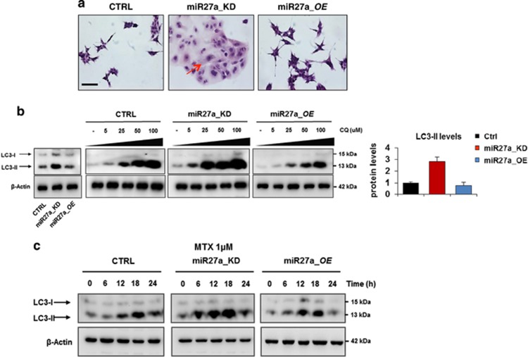
Figure 4.
miR-27a knockdown promotes autophagy in CRC cells. (a) Morphological features of HCT116 CRTL, miR27a_KD and miR27a_OE cells were evaluated by hematoxylin and eosin staining; the arrows point out some remarkable phenotypic characteristics (Scale bar, 20 mm). (b) Dose-dependent induction of autophagy in HCT116 CRTL, miR27a_KD and miR27a_OE cells exposed to the lysosomotropic drug chloroquine (CQ). Western blot analysis of the LC3-II mature form, an autophagic marker, is reported along with its relative quantification in the histogram. β-Actin was used as loading control. (c) Time-course of autophagy induction in HCT116 CRTL, miR27a_KD and miR27a_OE cells upon MTX treatment at 1 μM. Immunoblot of LC-3-II was used as a marker. All data are representative of three independent experiments and error bars represent S.D. of technical replicates (mean±S.D.)

