Figure 2.
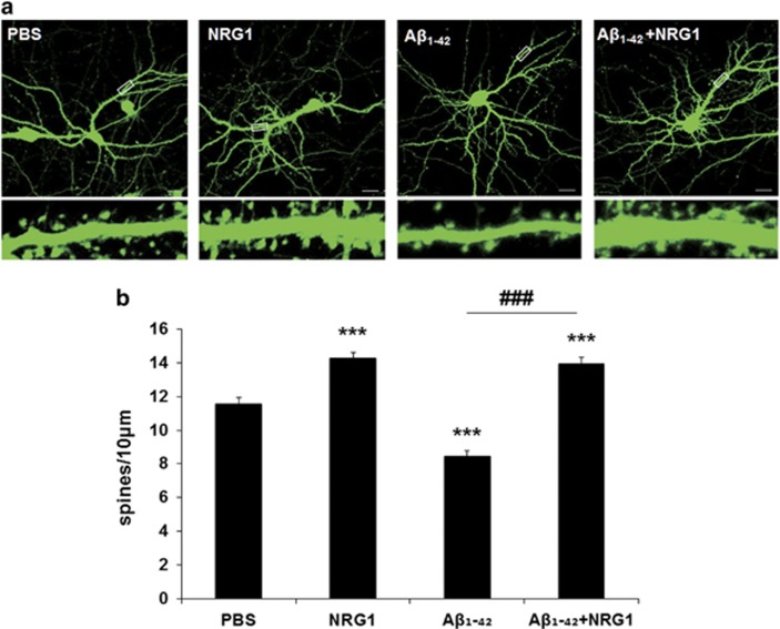
NRG1 rescues the decrease in dendritic spine density induced by oligomeric Aβ1-42 in rat primary hippocampal neurons. (a) Representative images of dendritic spines in primary hippocampal neurons at DIV 17 after treatment with oligomeric Aβ1-42, (at DIV 13) either alone or with PBS or NRG1 (at DIV 14) are shown. The outlined dendritic segment (upper) is enlarged to depict the spine morphology (bottom). Scale bars represent 20 μm (upper) and 5 μm (lower). (b) Quantification of spine density (40–80 μm of secondary dendritic spines from the soma) at DIV 17 after transfection with mGFP in primary hippocampal neurons at DIV 12. Treatment with 250 nM Aβ1-42 significantly decreased dendritic spine numbers, the number per 10 μm of dendrites (8.445±0.347, n=16, ***P<0.001), compared with PBS-treated controls (11.548±0.3875, n=18). Co-treatment with Aβ1-42 plus 10 nM NRG1 alleviated the decrease in dendritic spines induced by Aβ1-42 (13.960±0.367, n=19, ###P<0.001) compared with the Aβ1-42 treatment (8.445±0.347, n=16). Statistical analysis was performed via one-way ANOVA followed by Fisher's LSD post hoc test; the data are expressed as the mean±S.E.M.

