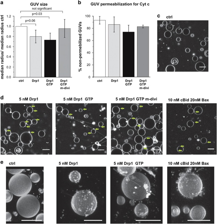
Figure 1.
Changes in GUV size upon incubation with Drp1. (a) Comparison of the median GUV radius from GUV incubated 90 min with only Cyt c488 (control) or additionally with 5 nM Drp1, 1 mM GTP or 50 μm mdivi-1 normalized to the median radius of the control sample. Notably, mdivi-1 partly precipitated, thus the effective mdivi-1 concentration is lower as 50 μM. Error bars in (a) and (b) correspond to the S.D. (N=3). (b) Percentage of non-permeabilized GUVs for Cyt c488 from (a). (c–e) Confocal images (c and d) and 3D reconstitution of z-stacks from GUV incubated with the size marker proteins alone or with the components indicated in the figure. In all experiments the GUVs were composed of a lipid mixture mimicking the MOM and labeled with <0.05% DiI. Green arrows in (d) indicate structures indicative for vesicle buds. Scale bar: 20 μm

