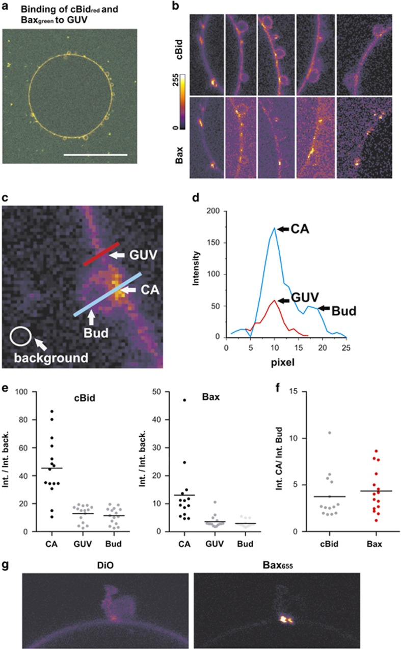
Figure 5.
cBid and Bax bind preferentially to highly curved membranes. (a) Image of one GUV composed of 30% CL and 70% PC after 30 min incubation with 20 nM cBid655 and 40 nM Bax488. Shown is the color merge. Scale bar: 50 μm. (b) Zoom-in's from (a) and other GUVs are shown to visualize that cBid and Bax bind preferentially at the highly curved interface between bud and GUV. A scale bar concerning the color code related to the pixel intensity is shown on the left side. (c) Example image on how the pixel intensities in the bud membrane, in the GUV membrane and in the contact area (indicated as CA) as well as the background are measured. The results are drawn in (d)–(f). (d) Display of the exemplary line scans shown in (c). (e) Pixel intensities for cBid and Bax at the contact area, the GUV and the bud (divided by the background intensity) from several GUV/bud pairs. (f) Ratio of the pixel intensity in the GUV/bud contact area compared with the pixel intensities at the GUV membranes. (g) Zoom-in from an exemplary GUV bud pair labeled with DiO and incubated with cBid unlabeled and 40 nM Bax655

