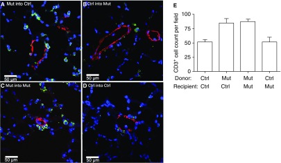
Figure 3.
Lungs of recipient mice transplanted with mutant (Mut) bone marrow (BM) cells had increased numbers of T cells (CD3+) compared with lungs of Mut mice transplanted with control (Ctrl) BM cells. Lungs of recipient mice were analyzed 16 weeks after transplantation by costaining with CD3–fluorescein isothiocyanate (green) and α-smooth muscle actin–tetramethylrhodamine (red) antibodies. Nuclei were visualized with 4′,6-diamidino-2-phenylindole (blue). (A) Mut BM transplanted into Ctrl recipient mice. (B) Ctrl BM transplanted into Mut recipient mice. (C) Control group; Mut BM cells transplanted into Mut recipient mice. (D) Control group; Ctrl BM cells transplanted into Ctrl recipient mice. Representative pictures are shown. (E) Average number of CD3+ cells by cell counting from 10 random fields at a magnification of ×10.

