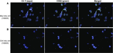
Figure 6.
Recipient mice contain donor-derived CD68+ cells. Lungs of female recipient mice transplanted with male bone marrow (BM) cells were analyzed 16 weeks after transplantation. To visualize the Y chromosome (Ch Y) in donor male BM cells, fluorescence in situ hybridization was combined with immunohistochemistry. Lung sections were painted with Y probe (aqua) and then costained with CD68–fluorescein isothiocyanate (macrophages, green). Nuclei were visualized with 4′,6-diamidino-2-phenylindole (blue). Representative pictures are shown at an original magnification of ×1,000. (A) Mutant (Mut) BM transplanted into control (Ctrl) recipient mice. (B) Control group; Ctrl BM transplanted into Ctrl recipient mice.

