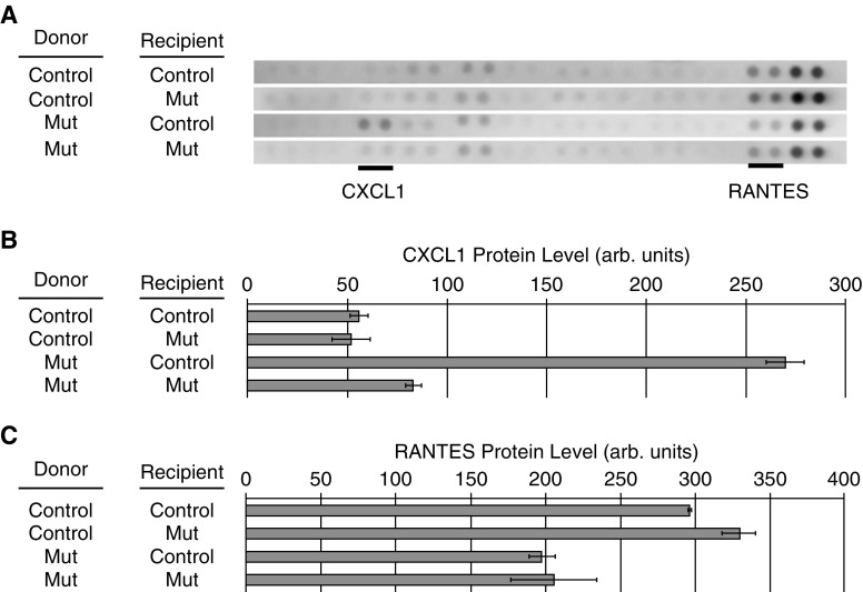
Figure 7.
Altered chemokine levels in the lungs of recipient mice. Protein extracts from lung tissue of three recipient mice from each transplant group were pooled and applied to R&D Systems mouse cytokine array panels. The four analysis groups were as follows: control (Ctrl) bone marrow (BM) transplanted into Ctrl recipient mice, Ctrl BM transplanted into mutant (Mut) recipient mice, Mut BM cells transplanted into Ctrl recipient mice, and Mut BM cells transplanted in Mut recipient mice. (A) Each pair of dots represents an independent cytokine antibody. Forty cytokines were analyzed per array. The strips containing CXCL1 and RANTES (regulated upon activation, normal T-cell expressed and secreted) are shown, and the spots representing CXCL1 and RANTES are underlined. (B) Normalized densitometry of CXCL1 in each group. (C) Normalized densitometry of RANTES in each group. arb. = arbitrary; CXCL1 = chemokine (C-X-C motif) ligand 1 (melanoma growth–stimulating activity α).

