Figure 5.
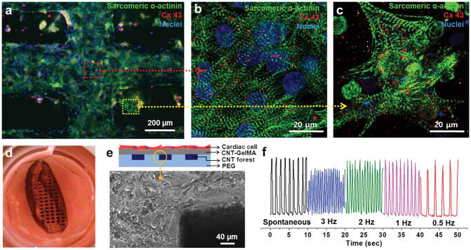
Cardiac tissue organization on composite hydrogel layers incorporating CNT forest electrodes. (a) Phenotype of cardiac cells. Immunostaining of sarcomeric α-actinin (green), nuclei (blue), and Cx-43 (red) revealed that cardiac tissues (5-day culture) were created on top of a multilayer hydrogel sheet impregnated with aligned CNT microelectrodes. Magnified image showing the well interconnected sarcomeric structures of cardiac tissue which is located (b) above locations between the electrodes (red dot arrow) and (c) above the electrodes (yellow dot arrow). (d) Photograph of a free-standing 3D bio-hybrid actuator cultured for 8 days. (e) Schematic illustration of a multilayer hydrogel sheet impregnated with aligned CNT microelectrodes (side view). Phase contrast image of the boundary between the CNT forest electrodes and the hydrogel layer shows that the cardiac tissues remained attached to the top hydrogel layer snd stayed intact. (f) The displacement of the CNT forest electrode in multilayer hydrogel sheets (yellow circled tip in b) over time under electrical stimulation (Square wave form, 1.2 V/cm, Frequency: 0.5 Hz – 3 Hz, 50ms pulse width).

