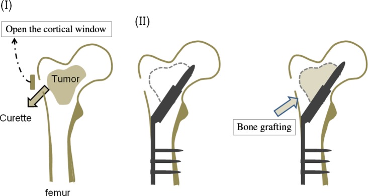
Figure 1.

This figure shows our surgical procedure. (I) A cortical window was made at the lateral femoral cortex. Through a cortical window, a biopsy sample was obtained for frozen section. The window must be large enough to allow adequate curettage of the tumor until underlying normal bone is exposed. (II) After the lateral cortex and femoral head were reamed along with a guide wire which was inserted under radiological control, a compression hip screw was inserted. (III) The synthetic bone was implanted in the bone defect.
