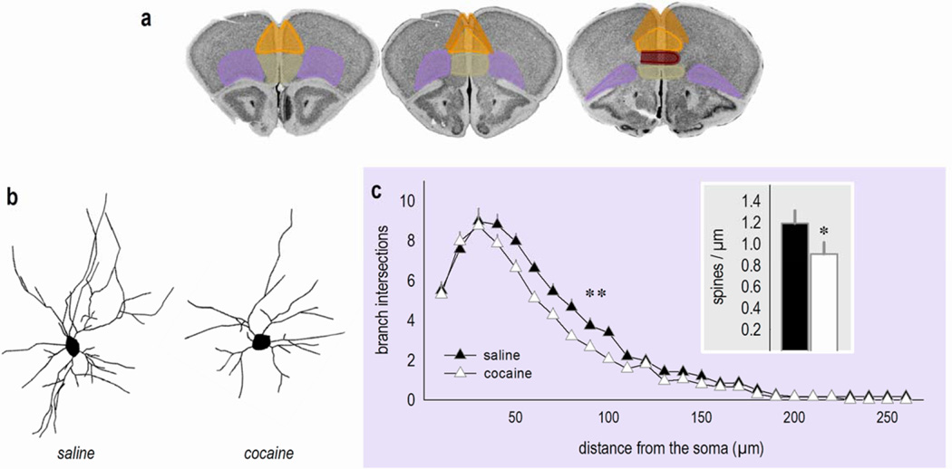
Figure 1. Cocaine: A double threat to neurons in the oPFC?
(a) Sub-regions of the prefrontal cortex transposed onto coronal images from the Mouse Brain Library (202). Pink represents the oPFC and orange, yellow, red and green represent sub-regions of the mPFC: the anterior cingulate, prelimbic, infralimbic, and medial orbital cortices, respectively. These sections correspond roughly to Bregma +1.98, +2.1, and +2.46. (b) Representative deep-layer oPFC neurons from mice treated with saline or cocaine several weeks prior to euthanasia. (c) Sholl analyses indicate that cocaine exposure decreases branch intersections. Inset: Dendritic spines on secondary and tertiary branches are also lost. These findings were originally reported in ref. 81 (for dendrites) and ref. 71 (for dendritic spines), and the reader is referred to these reports for methodological details. Bars and symbols represent means and SEMs, *p<0.05,**p<0.05 40–100 µm from the soma.

