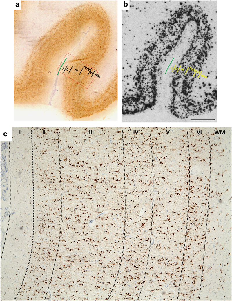
Figure 1.
Representative images showing cortical lamina by (a) immunostaining (NeuN) and (b) in situ hybridization (somatostatin) in the orbital frontal cortex. (c) Orbitofrontal cortical lamina identification (layers I–VI) based on NeuN immunostaining (magnification ×4). Red rectangular box in a represents the cortical area shown in c.

