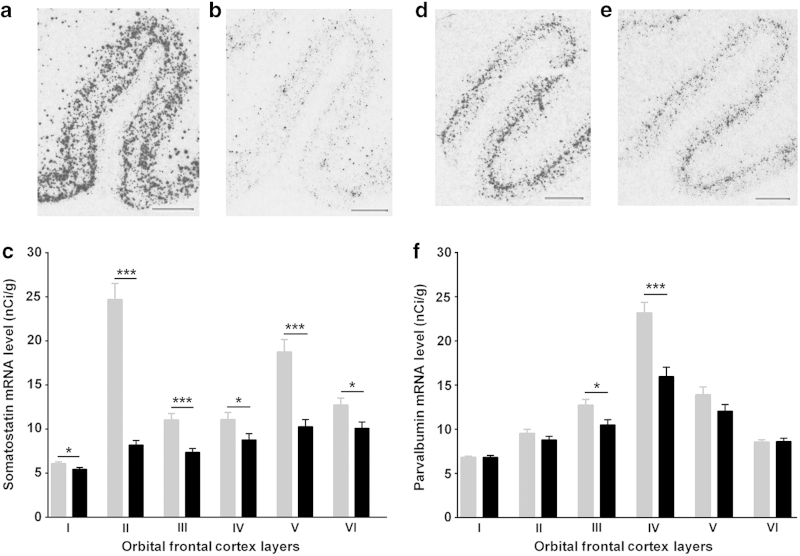
Figure 3.
Representative in situ hybridization images showing somatostatin (SST: a, b) and parvalbumin (PV: d, e) mRNA distributions in human orbital frontal cortex in control (a, d) and schizophrenia (b, e) subjects, respectively. Laminar-specific SST (c) and PV (f) mRNA levels (nCi/g) in the gray matter in control (gray) and schizophrenia (black) subjects (*P<0.05; ***P<0.001). Scale bar=2 mm; error bars represent s.e.m.

