Figure 1.
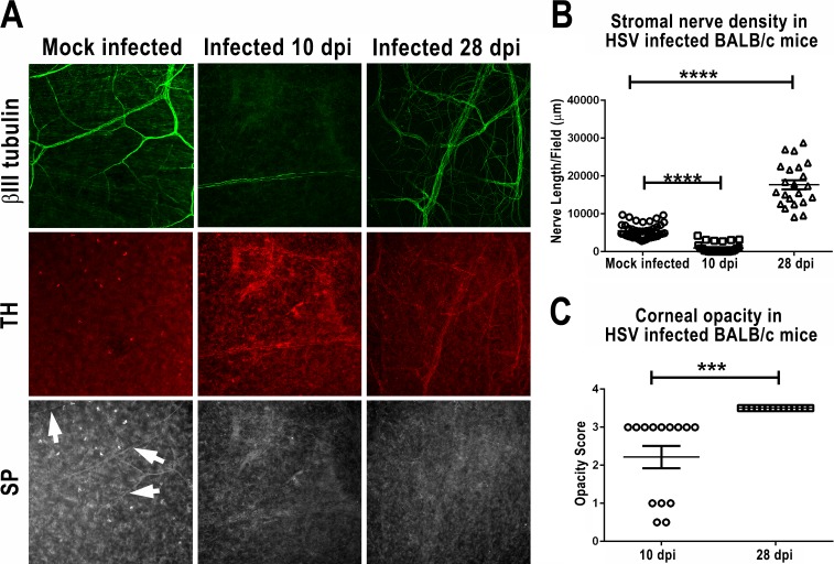
BALB/c mice developed sympathetic nerve hyperinnervation associated with severe HSK at 28 dpi. BALB/c mice were mock infected or infected with 1 × 10⁵ pfu of HSV-1 KOS strain. At 10 or 28 dpi, the corneal opacity was recorded, and then the mice were killed, infected corneas were excised and fixed, and whole mounts were stained for the neuronal marker βIII tubulin (green), the sympathetic nerve marker TH (red), and the sensory nerve marker SP (gray). Confocal images were acquired and analyzed as described in Methods. (A) Changes in nerve innervation of the stroma at 10 and 28 dpi. The corneal stroma of mock infected mice show a low density of nerves that express SP but not TH. In contrast, corneas obtained at 10 dpi exhibit an almost complete lack of corneal nerves, whereas corneas obtained at 28 dpi show a corneal stroma that is hyperinnervated by nerve fibers that express the sympathetic marker TH but not the sensory marker SP. (B) Stromal nerve density measured by cumulative length of nerve fiber at 10 or 28 dpi. (C) Corneal opacity in HSV-infected mice recorded prior to death at 10 or 28 dpi. No opacity was observed in mock infected corneas (data not shown). ***P < 0.001, ****P < 0.0001.

