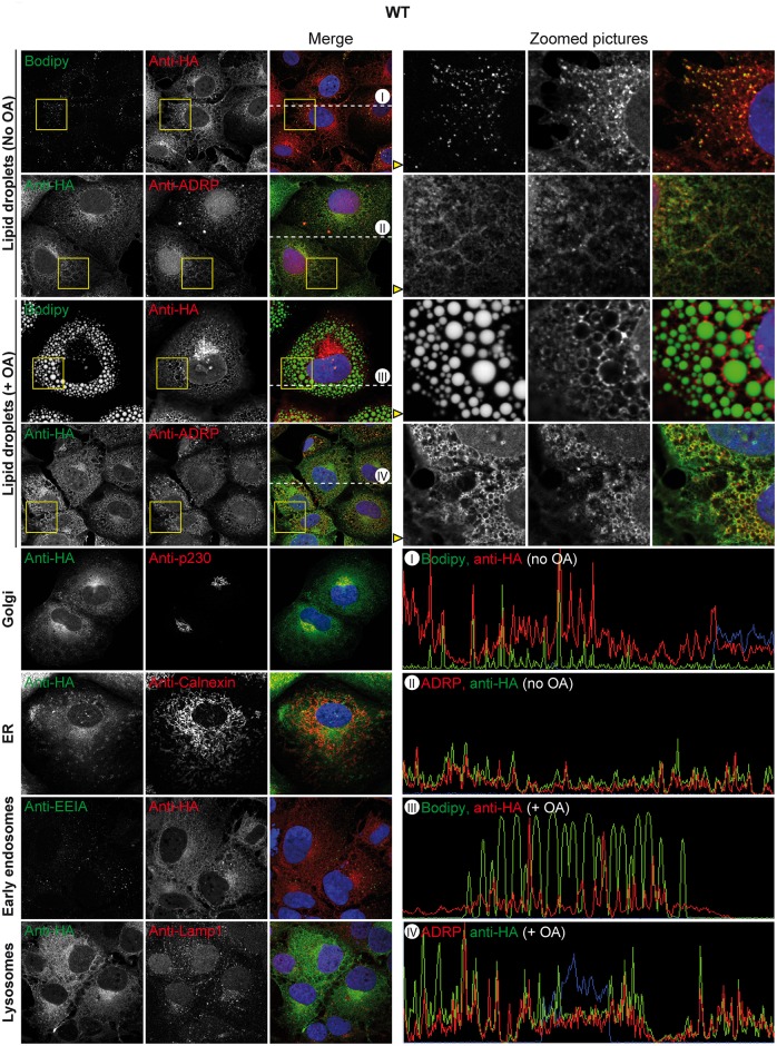
Fig 3. Subcellular localisation of ABHD5.
Lunet N hCD81 cells expressing HA-tagged ABHD5 were stained for the HA epitope as well as for diverse organelles (LD (Bodipy or ADRP), trans-Golgi (p230), ER (calnexin), early endosomes (EEIA), lysosomes (Lamp1)) and counterstained with Dapi. In each panel, representative pictures are shown on the left part. For the colocalisation analysis with the lipid droplet markers Bodipy and ADRP, a portion of the image highlighted with a yellow square is magnified on the right side and depicted in the different channels in the same order. Moreover, for the same pictures, the intensity profile of the different dyes along the dotted line in the merged picture is shown in the lower right quarter and labelled with a Roman number.

