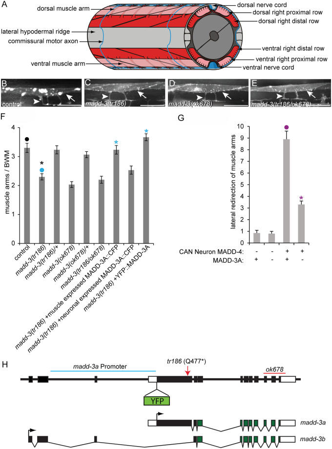
Fig 1. The muscle arm extension defects of madd-3 mutants.
A. A schematic of muscle arms and their motor neuron targets. B-E. The muscle arms of animals of the indicated genotype. All animals harbour the trIs30 transgene. Scale bars are 25μm. F. The number of muscle arms that extend from muscle VL11 towards the ventral nerve cord. G. The number of muscle arms that extend towards MADD-4 that is ectopically expressed by the lateral CAN neuron. Numbers shown are for the left side of the animal. The right side behaves similarly. In both graphs an asterisks indicates a significant difference (p<0.01) compared to the data point indicated with a closed circle with the same color as the asterisks. H. Schematics of the genomic madd-3 locus and resulting transcripts. The position of the tr186 allele and the ok678 deletion are indicated. The position where a YFP tag was inserted in the context of the fosmid reporter (see text) is shown. The region of DNA used to drive the madd-3a YFP transcriptional reporter is indicated with a blue line. Black boxes indicate exons and white boxes are untranslated regions.

