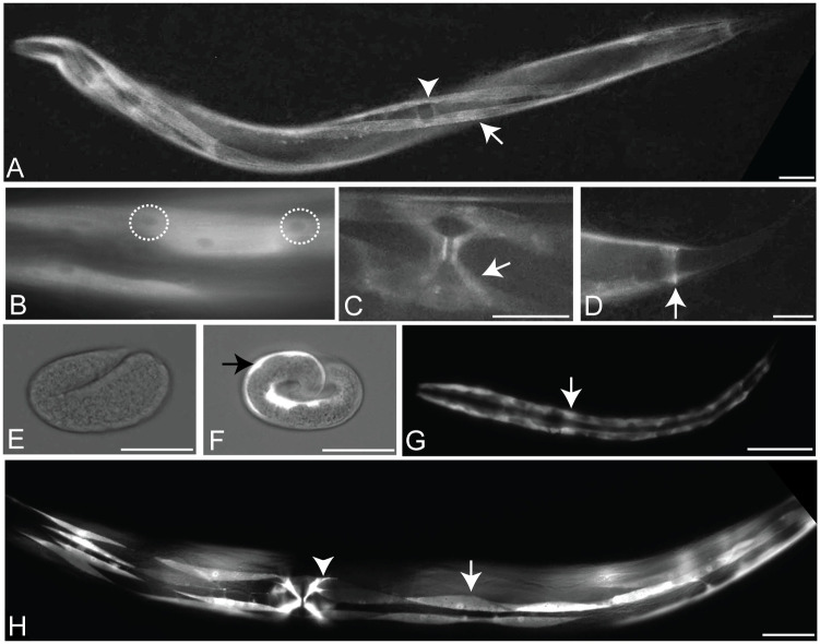
Fig 2. The MADD-3 expression pattern.
A-D. Expression of a MADD-3A translational reporter (the placement of the YFP tag is shown in Fig 1H). A. A young adult worm. An arrow indicates a body wall muscle within one of the ventral quadrants, the arrowhead indicates the position of the vulval slit. The animal is twisted due to a marker within the extra-chromosomal array that carries the reporter. B. Body wall muscles showing relatively less YFP in the nucleus (two nuclei are encircled). C. The vulva muscles (arrow). D. The anal depressor muscle (arrow). E-H. Expression of a madd-3Ap::YFP transcriptional reporter. E. A partial bright-field photograph showing that YFP is not expressed from the madd-3A promoter at the embryonic two fold stage. F. At the embryonic pretzel stage, YFP expression driven by the madd-3A promoter is evident in the body wall muscles (arrow). G. An L1 larva with the two rows of body wall muscles showing. H. A young adult worm. A single body wall muscle is indicated with an arrow and a vulva muscle is indicated with an arrowhead. Scale bars represent 25 μm.

