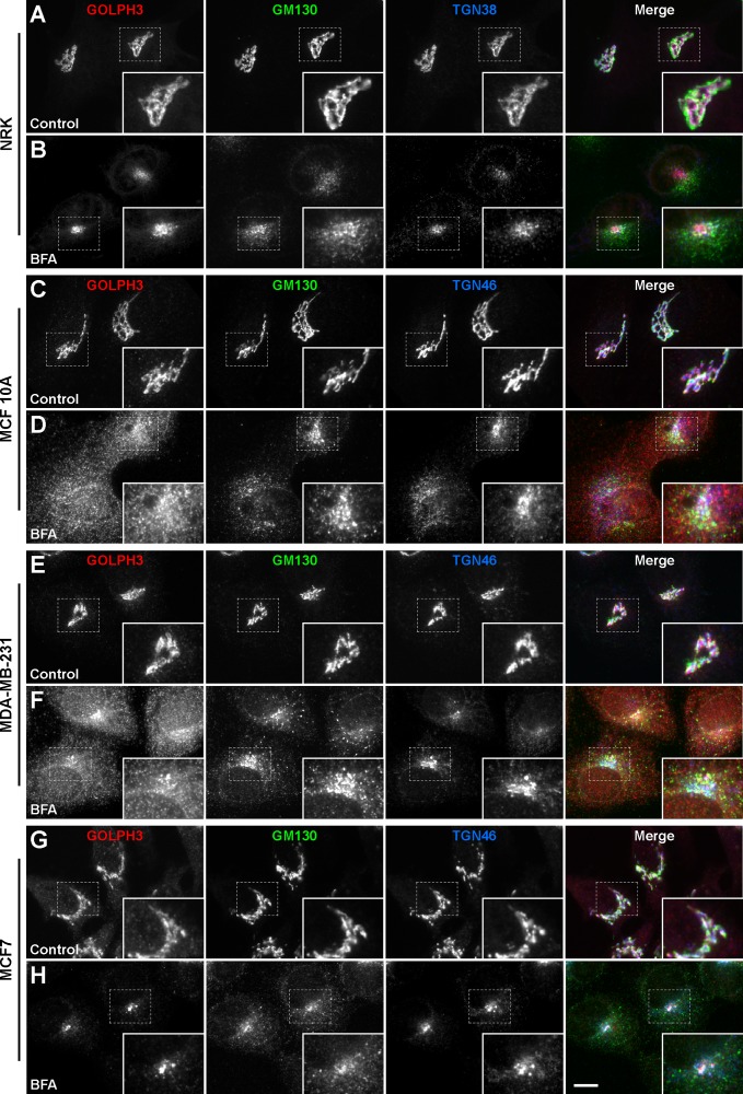
Fig 4. The sensitivity of GOLPH3 to BFA is different in different human breast cell lines.
NRK (A and B), MCF 10A (C and D), MDA-MB-231 (E and F), and MCF7 (G and H) cells were left untreated (Control) or treated with 5 μg/ml BFA for 60 min (BFA). Cells were fixed, permeabilized, and immunolabeled with rabbit polyclonal antibody to GOLPH3, mouse monoclonal antibody to GM130, and either sheep antibody to TGN38 (A and B) or sheep antibody to TGN46 (C to H). Secondary antibodies were Alexa-594-conjugated donkey anti-rabbit IgG (red channel), Alexa-488-conjugated donkey anti-mouse IgG (green channel), and Alexa-647-conjugated donkey anti-sheep IgG (blue channel). Stained cells were examined by fluorescence microscopy. Merging red, green, and blue channels generated the fourth image on each row; yellow indicates overlapping localization of the red and green channels, cyan indicates overlapping localization of the green and blue channels, magenta indicates overlapping localization of the red and blue channels, and white indicates overlapping localization of all three channels. Insets show 1.7x magnifications. Bar, 10 μm.

