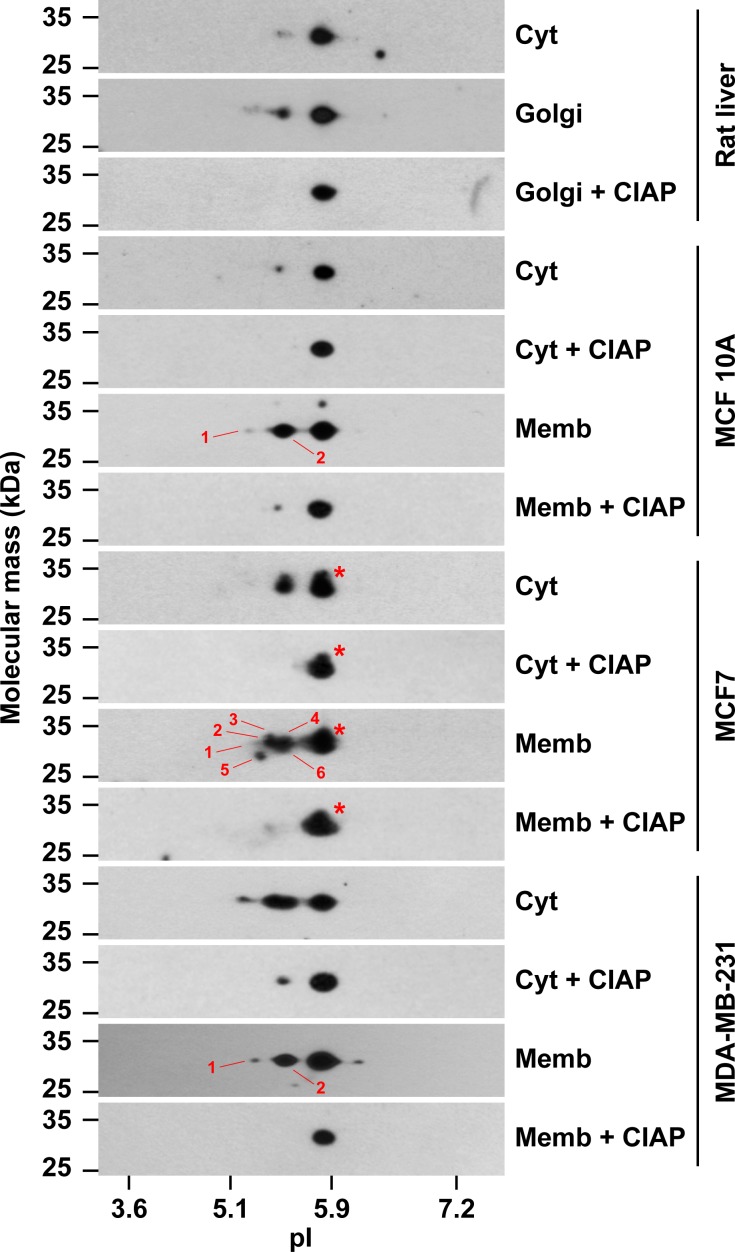
Fig 7. The cytosolic and membrane pools of GOLPH3 are differentially modified in different human breast cell lines.
Samples (30 μg of proteins) of rat liver cytosol (Cyt), rat liver Golgi membranes, and of cytosolic (Cyt) and membrane (Memb) fractions from the cell lines indicated at the right were analyzed by two-dimensional gel electrophoresis (2-D GE) and immunoblotting using antibody to GOLPH3. Samples of rat liver Golgi membranes, and of the cytosolic and membrane fractions of each cell line, were dephosphorylated with calf intestine alkaline phosphatase (CIAP) before processing for 2-D GE. The position of molecular mass markers is indicated on the left. The position of isoelectric point (pI) markers is indicated at the bottom. Red asterisks indicate the position of additional, less abundant, but distinct spots in the samples of MCF7 cells that have slightly slower electrophoretic mobility. Numbers indicate different acidic forms identified in immunoblot films subjected to different exposure times.

