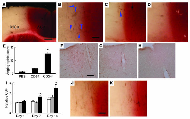
Figure 2.
Transplantation of CD34+ cells after stroke accelerates neovascularization. (A–D) Mice subjected to stroke received CD34+ cells (A and B), CD34– cells (C), or PBS alone (D) on day 2. Animals were infused with carbon black ink and killed at 24 hours after cell transplantations. Sections were stained with TTC. Neovascularization was noted at the border zone between the ACA and MCA areas (arrowheads show microvessels), especially in animals treated with CD34+ cells compared with those receiving CD34– cells or PBS alone. (E) An angiographic score for each experimental condition based on analysis of 6 mice per group. (F–H) Activated endothelial cells were observed with antibody specific for mouse CD13 in the ACA area. F: CD34+ cells; G: CD34– cells; H: PBS. (I) CBF was measured in the MCA area just outside of the penumbra, and values in animals treated with CD34+ cells (black bars), CD34– cells (gray bars), or PBS (white bars) were compared with values before cell transplantation at times corresponding to days 1, 7, and 14 after cell transplantation (n = 6 per group). Data shown are relative CBF versus day the measurement was performed. (J and K) Labeling vasculature by infusion of carbon black ink demonstrated neovasculature at the border of the MCA and ACA cortex in animals treated with CD34+ cells (J) and CD34+/Flk-1– cells (K) on day 7 after cell transplantation. Scale bars: 0.5 mm (A) and 0.1 mm (B, F, and J). *P < 0.05 versus PBS.

