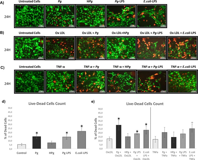
Fig 2. Qualitative evaluation of the EC death.
(A) Viability of HUVECs infected with Pg at a MOI of 100 or Heat-inactivated Pg (HPg) and stimulated by Pg-LPS (1μg/ml) or E.Coli-LPS (1μg/ml) at 24h. All different conditions have been evaluated quantitatively and qualitatively by Live-Dead staining assays. (B) Viability of Ox-LDL (50μg/ml) pre-treated HUVECs on cell cultures infected with Pg or HPg at a MOI of 100 and stimulated by Pg-LPS (1μg/ml) or E.Coli-LPS (1μg/ml) at 24h. (C) Viability of TNF- α (10ng/ml) pre-treated HUVECs on cell cultures infected with Pg or HPg at a MOI of 100 and stimulated by Pg-LPS (1μg/ml) or E.Coli-LPS (1μg/ml) at 24h. (D) Percentage of dead cells infected with Pg or HPg and stimulated by Pg-LPS (1μg/ml) or E.Coli-LPS (1μg/ml) at 24h. (E) Percentage of dead cells in OxLDL qnd TNF- α pre-treated HUVECs. All images were acquired under fluorescence microscopy (in green: viable cells; in red: dead cells). All scale bars indicate 100 μm.

