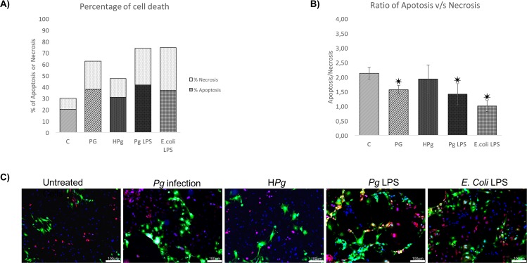
Fig 3. Infection of ECs leads to cell death mediated by apoptosis.
(A) Percentage of cell death of ECs infected with Pg or Heat inactivated Pg (HPg) at a MOI of 100 and stimulated by Pg-LPS (1μg/ml) or E.Coli-LPS (1μg/ml). Each percentage was calculated on account of total cells counted in triplicate for each experiment. (B) The apoptosis/necrosis ratio of ECs infected with Pg or Heat inactivated Pg (HPg) at a MOI of 100 and stimulated by Pg-LPS (1μg/ml) or E.Coli-LPS (1μg/ml). Each value was calculated from the ratio between the total number of apoptotic cells and necrotic cells and for each count nine images were used of each experimentation. Data were expressed as mean ± SD. ✷: difference between non pre-treated/stimulated/infected and infected/stimulated cells, p < 0.05; (C) Infected and stimulated HUVECs cell death was evaluated for each condition qualitatively using Annexin V-IP staining at 24h (in green: Annexin V positive staining; in red: Iodure propidium positive staining; in blue: DAPI nuclear staining) Images were acquired under fluorescence microscopy (10x) after Annexin V-IP and DAPI staining for all previously described condition. All scale bars indicate 100 μm.

