Figure 1.
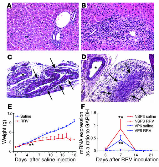
RRV infection induces biliary inflammation and growth failure in neonatal mice. WT Balb/c mice were injected with normal saline (control) or RRV within 24 hours of birth, and the hepatobiliary system was examined 7 days later. (A) While livers of control mice had normal appearance of the portal tracts, RRV challenge resulted in the expansion of portal spaces by inflammatory cells and proliferating bile duct cells (B). (C) Cross section of the extrahepatic bile duct of a control mouse revealed normal epithelium and unobstructed lumen (arrows). (D) In contrast, injection of RRV produced lumenal obstruction of extrahepatic bile ducts (arrows). Tissue sections were stained with H&E. Magnification of ×400 for A and B, ×200 for C and D. Single asterisks denote neighboring arteries in C and D. (E) It can be seen that RRV injection also led to poor growth during the suckling period. **P < 0.01 when compared with controls at days 7–16; n = 25 mice in the beginning of the experiment. Expression of mRNA encoding RRV nonstructural (NSP3) and structural (VP6) proteins was high at day 7 but (F) undetectable at day 14. **P < 0.01; n = 4–7 mice per group at each time point.

