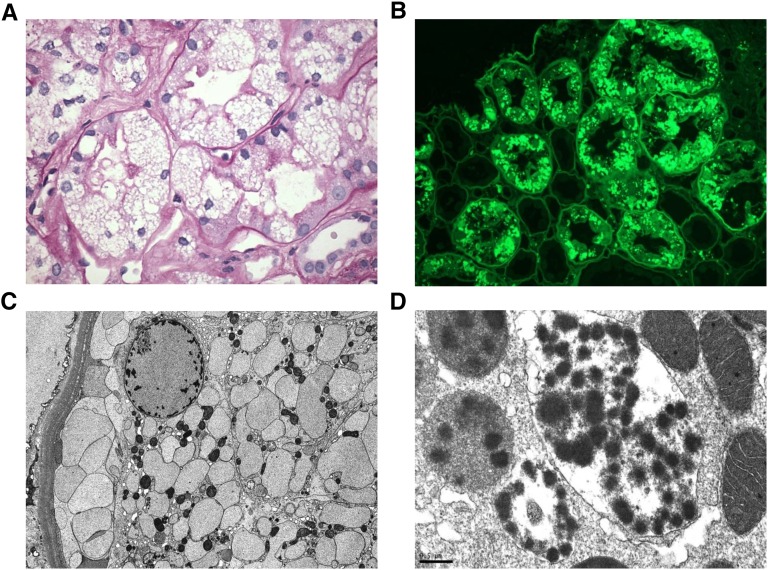
Figure 2.
Pathologic features of noncrystalline LCPT. (A) The proximal tubular cells are variably distended by abundant PAS-negative vacuoles associated with focal loss of brush border (PAS, ×600). (B) By immunofluorescence performed on pronase-digested paraffin sections, the vacuoles stain intensely for κ light chain (FITC-conjugated antisera to κ light chain, ×400). (C) By EM, the vacuoles appear rounded or ovoid and contain finely granular material without crystal formation. The numerous membrane-bound vesicles extend from the apical to the basal cytoplasm and crowd out the mitochondria, which appear reduced in size and number (Magnification, ×5000). (D) A different case of noncrystalline LCPT exhibits membrane-bound phagolysosomes with a mottled appearance owing to rounded electron-dense particulate inclusions suspended free on an electron-lucent background or within a moderately electron-dense amorphous matrix. The adjacent mitochondria appear well preserved (Magnification, ×40,000).

