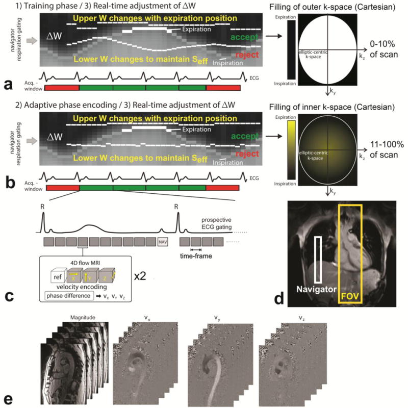
Figure 1.

Data acquisition with prospective ECG gating and respiratory navigator gating consisting of (a) a training phase for the initial adjustment of the navigator acceptance window (ΔW) during the collection of the initial 10% of the total 4D flow data during acquisition of outer ky–kz-space and (b) continued real time adjustment of ΔW in combination with respiratory driven phase encoding for the remaining 90% of data acquisition. (c) The 4D flow scan was prospectively ECG gated and for each time-frame, four datasets, one reference scan and three flow-sensitive scans, were acquired in an interleaved fashion. (d) The navigator was placed on the lung-liver interface, and the field of view (FOV) covered the entire thoracic aorta. (e) The resulting 4D flow data consisted of time resolved 3D magnitude data, and three time-resolved 3D phase difference datasets representing blood flow velocity in x, y and z-direction.
