
Figure 4.
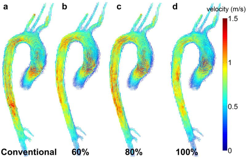
Examples (volunteer 7, 3T) for 3D velocity vector images at peak systole for (a) conventional navigator (scan 1) gating and for the new navigator scheme (scans 2–4) with fixed Seff = (b) 60%, (c) 80% and (d) 100%.
Official websites use .gov
A
.gov website belongs to an official
government organization in the United States.
Secure .gov websites use HTTPS
A lock (
) or https:// means you've safely
connected to the .gov website. Share sensitive
information only on official, secure websites.
Examples (volunteer 7, 3T) for 3D velocity vector images at peak systole for (a) conventional navigator (scan 1) gating and for the new navigator scheme (scans 2–4) with fixed Seff = (b) 60%, (c) 80% and (d) 100%.