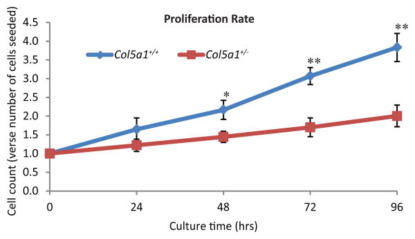
Figure 2. EDS dermal fibroblasts proliferated slower than wild type controls.
Representative data demonstrating decreased proliferation of EDS dermal fibroblasts. Proliferation rate was calculated as ratio of cell count at 24, 48, 72 and 96hrs versus number of cells plated (0 hour cell number). There was no significant difference in proliferation at 24 hr. A significant decrease in fibroblast proliferation was observed in Col5a1+/- fibroblasts at 48hrs (*p<0.05), 72hrs and 96hrs (**p<0.01) relative to the Col5a1+/+ fibroblasts. Differences in the proliferation rate from 24 to 96 hrs were seen as a decrease in the slope for the Col5a1+/- fibroblasts. The experiments were repeated 3 times with 3 strains derived from Col5a1+/+ and Col5a1+/- dermis.

