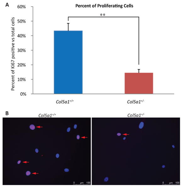
Figure 3. Decreased proliferation marker Ki-67 in EDS compared with wild type dermal fibroblasts.

Representative data demonstrating that proliferating Col5a1+/- fibroblasts were significantly decreased compared to Col5a1+/+ fibroblasts. Fibroblasts were incubated at 37°C for 24hr, fixed and labeled with anti-Ki67 antibody. Proliferating cells were detected by anti-Ki67, and nuclei were counterstained with DAPI. Number of Ki67 positive cells and total cells were counted. (A) Percentage of proliferating Col5a1+/- cells (14.5%) was ∼ 30% of that of Col5a1+/+ cells (43.3%) (**p<0.01). Ratio was calculated by number of Ki67 positive cells versus total number of cells. (B) Representative immunofluorescence micrographs showing decreased proliferation in Col5a1+/- compared with Col5a1+/+ fibroblasts. Proliferating fibroblasts were identified using anti-Ki67 (arrows). Anti-Ki-67 (red), nuclear localization with DAPI (blue). The experiments were repeated 3 times with 3 strains derived from Col5a1+/+ and Col5a1+/- dermis.
