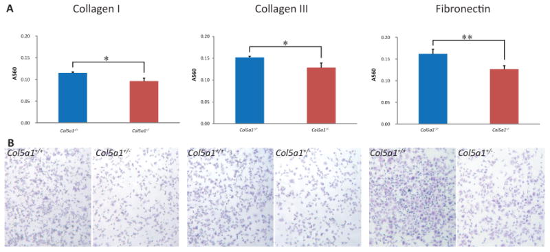
Figure 4. Decreased attachment to wound matrix components in EDS compared with wild type fibroblasts.

Representative analysis of fibroblast attachment. Fibroblasts were seeded on plates coated with collagen I, collagen III or fibronectin and incubated for 2hrs. Plates were washed and attached cells were stained using Crystal Violet. (A) The dye was extracted and the absorbance was measured at 560nm (A560). A560 values showed that more Col5a1+/+ fibroblasts attached compared to Col5a1+/- fibroblasts at a level of significance of p<0.05(*) on collagen I and collagen III, and p<0.01 (**) on fibronectin at 2hr. (B) Attachment of Col5a1+/- and Col5a1+/+ fibroblasts on collagen I, collagen III, and fibronectin were shown under light microscopy (4×). Attached fibroblasts were visualized in blue color by staining with Crystal Violet. The experiments were repeated 3 times with 3 strains derived from Col5a1+/+ and Col5a1+/- dermis.
