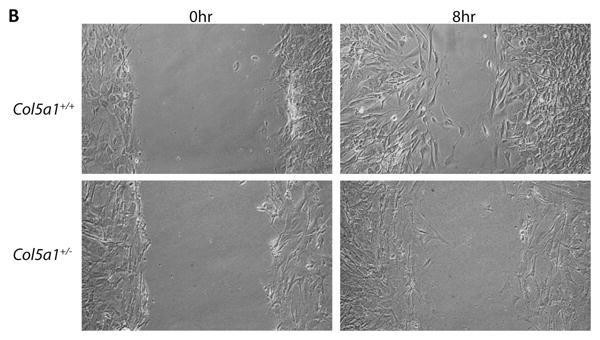
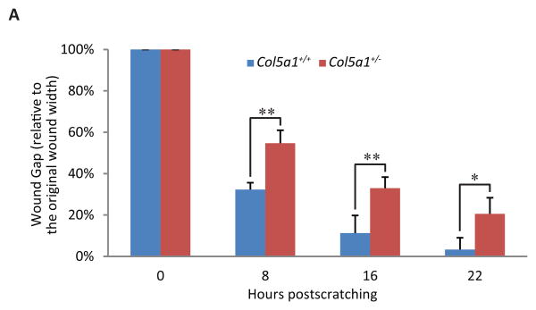
Figure 5. Fibroblast migration and in vitro wound closure is reduced in EDS compared to wild type dermal fibroblasts.

A representative analysis of in vitro scratch wound closure showed a significant difference between Col5a1+/- fibroblasts and Col5a1+/+ fibroblasts. A scratch wound was created on the monolayer of Col5a1+/- and Col5a1+/+ fibroblasts. Images were acquired after culture at 37°C for 0, 8, 16, 22 and 30hrs after scratching and the wound gap width was measured. (A) Wound closure was expressed as percentage of wound gap width relative to the scratch width (shown as 0 hour width). There was a significant difference in closure at all time points analyzed: 8hr and 16hr (**p<0.01) and 22hr (*p<0.05). (B) Representative wound closure of Col5a1+/- and Col5a1+/+ fibroblasts at 0hr and 8hr are shown. The experiments were repeated 3 times with 3 strains derived from Col5a1+/+ and Col5a1+/- dermis.

