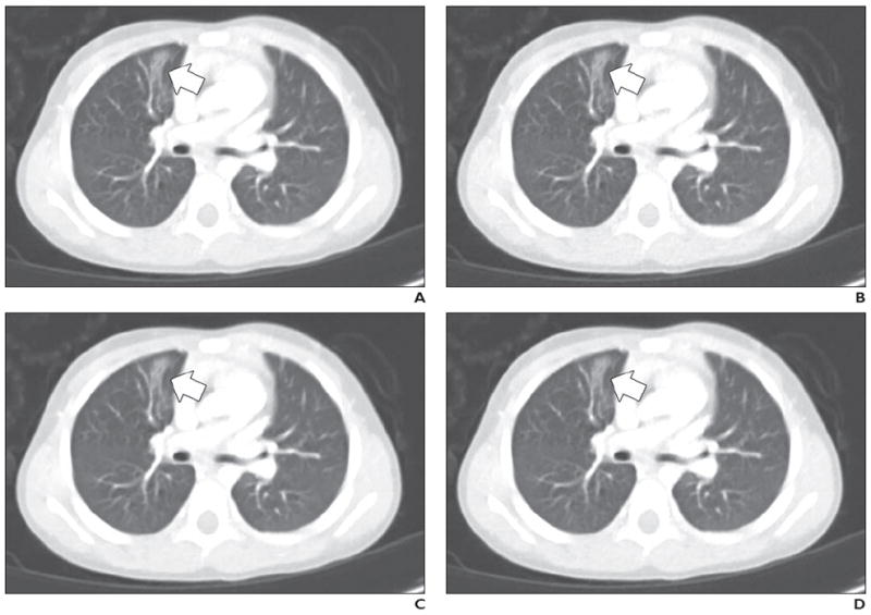
Fig. 5.

3-year-old girl with atelectasis in right upper lobe medially (arrow).
A–D, Contrast-enhanced chest CT images shown compare four dose plus reconstruction settings: full dose plus filtered back-projection (FBP) (A), half dose plus FBP (B), half dose plus sinogram-affirmed iterative reconstruction (SAFIRE, Siemens Healthcare) (C), and half dose plus adaptive nonlocal means (aNLM) (D). Scanning parameters included kilovoltage of 100 kV and full-dose volume CT dose index of 2.2 mGy. Half dose was 1.1 mGy. Average scores from all three readers for four images were as follows: 3.7 for full dose plus FBP, 3.0 for half dose plus FBP, 3.7 for half dose plus SAFIRE, and 4.3 for half dose plus aNLM.
