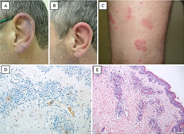
Figure 1. Skin lesions: A, Retiform purpura with a small area of necrosis in the right earlobe (pre-treatment). B, Residual skin lesions in the right earlobe after 3 weeks of immunosuppressive treatment. C, Purpuric violaceous lesions with surrounding erythema in the lower limb. Skin biopsy: D, Immunohistochemistry with anti-CD61 antibody showing positive staining for thrombi inside the vascular lumen, with surrounding inflammation of the vessel wall (magnification 100×). E, Small vessel vasculitis with neutrophilic inflammation and leukocytoclasia (H&E, magnification 100×).