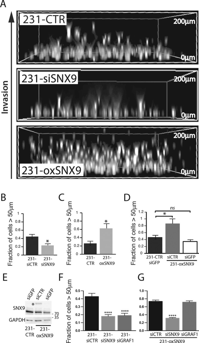
FIGURE 3:

SNX9 regulates the ability of MDA-MB-231 cells to invade through collagen matrix. (A) 231-CTR, -siSNX9, or -oxSNX9 cells were subjected to an inverted three-dimensional cell invasion assay through a bovine collagen I matrix (see Materials and Methods). Representative images of the positions of the nuclei of invading cells detected by Hoechst staining under the indicated conditions. (B, C) Quantification of nuclei distribution in inverted invasion assay of control cells vs. 231-siSNX9 (B) or 231-oxSNX9 (C). n = 10 and 6, respectively; *p = 0.02. (D) Quantification of cell invasion after specific depletion of exogenous SNX9, using an siGFP treatment of 231-oxSNX9. n = 4; *p = 0.04; ns, nonsignificant. (E) Western blot analysis of SNX9 expression in cell lines used in D. GAPDH was used as loading control. Blot is representative of three independent experiments. (F, G) Quantification of cell invasion of siRNA-treated parental MDA-MB-231 (F) or in siSNX9- or siGRAF1-treated 231-oxSNX9 (G). n = 3; ****p < 0.0001.
