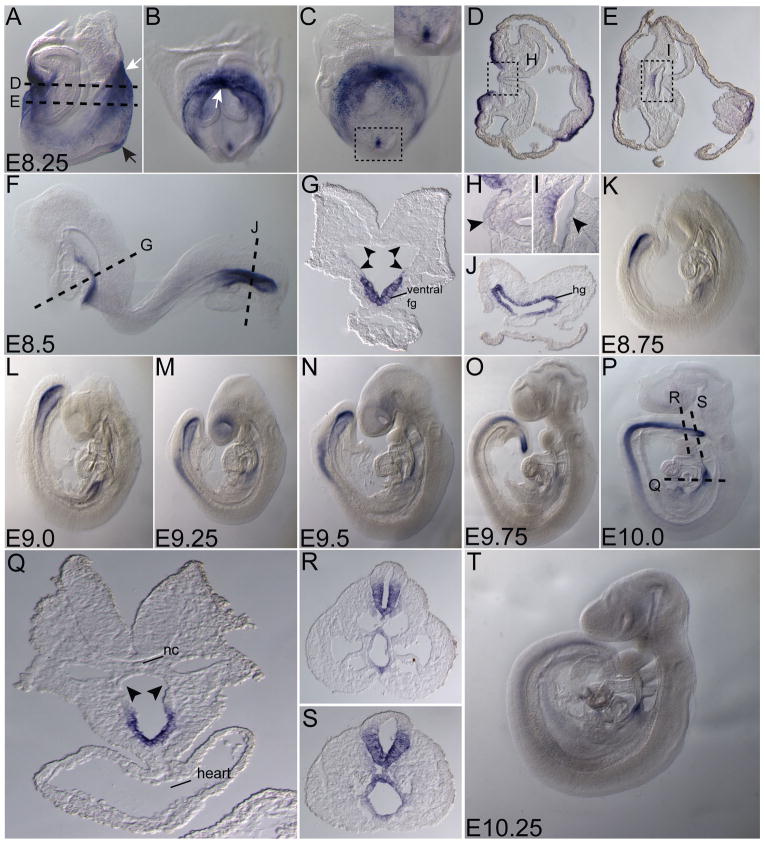
Figure 3. Expression of Ende in the definitive endoderm and neural tissue.
Lateral views (A, F, K–P, T), frontal view (B), posterior view (C) and transverse sections (D, E, G–J, Q–S) of representative embryos expressing Ende at the indicated stages. At E8.25, Ende expression was enriched in the foregut pocket and the presumptive hindgut of the embryo (white arrows in A, B). Expression in the ectoderm at the distal part of the embryo was maintained (black arrow in A), and was absent from the dorsal midline and flanking cells of the developing foregut (D, E; arrowheads in H, I). At E8.5, Ende expression continued to be specific to the ventral aspect of the foregut and absent in the remainder of the foregut (F; arrowheads in G), while expression was present throughout the hindgut (F, J). By E9.25 cells of the neural tube began to express Ende. As the embryo developed towards E10.0 Ende expression remained in the foregut overlying the heart, absent in the remainder of the foregut (P–S; arrowheads in Q), and was expressed in ventral part of the neural tube in the posterior half of the embryo (N–P, R, S). As late as E10.25 expression in the neural tube, hindgut and foregut were observed at reduced levels was significantly reduced (T). Foregut (fg), hindgut (hg), notochord (nc), embryonic day (E). E8.0 (1–4 somites), E8.5 (8–12 somites)

