FIG. 1.
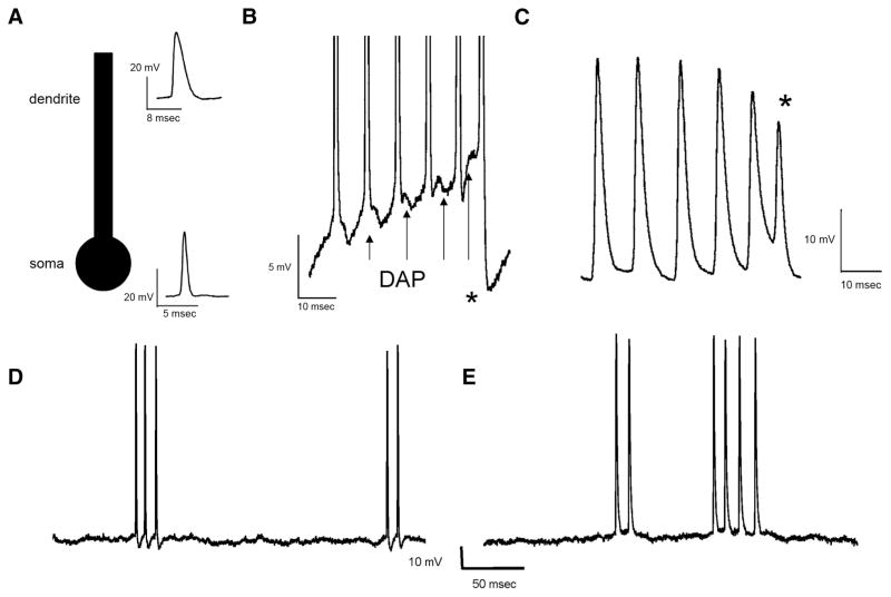
Differences between bursting in vivo and in vitro. A: schematic representation of the somatic and dendritic compartments that are necessary for bursting seen in vitro. Insets: a dendritic action potential (top) and somatic action potential (bottom). B: somatic in vitro recording from a pyramidal cell showing the ramp depolarization during the burst, the depolarizing afterpotential (DAP) growth (arrows), and the shortening of the interspike interval. The burst terminates with a burst afterhyperpolarization (bAHP, asterisk). C: dendritic in vitro recording from a pyramidal cell showing the decrease in spike height throughout the burst that is terminated by a dendritic failure (asterisk) when the interspike interval falls below the dendritic refractory period. D and E: example in vivo recordings from pyramidal cells. Based on spike shape, D resembles an in vitro somatic recording, whereas E resembles an in vitro dendritic recording. However, we found no evidence for depolarizing ramps, bAHPs, dendritic failures, or a decrease in spike height during the burst.

