Figure 3.
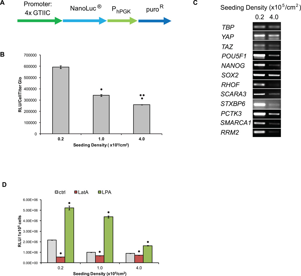
YAP transcriptional activity decreased as hPSC density increased. (A) Schematic of YAP transcriptional reporter construct. 4xGTIIC are 4 repeats of TEAD binding sites (ACATTCCA) that drive expression of NanoLuc. (B) hPSCs were singularized and plated at densities of 0.2 to 4.0 × 10⁵ cell/cm2. After 3 days in culture, 100,000 cells were harvested for luciferase assays. Chemiluminescent signal was normalized to CellTiter-Glo signal. Error bars represent standard deviation. (• indicates p<0.05 compared to 0.2 ×10⁵ cell/cm2 seeding density, •• indicates p<0.05 compared to 1.0 ×10⁵ cell/cm2 seeding density) (C) After 3 days, RNA was extracted for end-point RT-PCR analysis of YAP target genes with TATA-box binding protein (TBP) expression serving as the housekeeping control. YAP, TAZ, POU5F1 (Oct4), NANOG and SOX2 expression were also analyzed. (D) H9 4xGTIIC-Nluc cells were singularized and plated at densities of 0.2 to 4.0 × 10⁵ cell/cm2. The following day, cells were treated with 1 µM LatA or 2 µM LPA. After 24 hours, cells were harvested for luciferase assays and the chemiluminescent signal was normalized to cell count. Error bars represent standard deviation. (• indicates p<0.05 compared to no treatment condition at the same seeding density)

