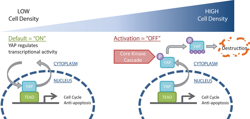
Figure 6.
Schematic model of the effects of hPSC density on YAP signaling. At low cell density (left), YAP is primarily localized to the nucleus where it associates with TEAD to regulate gene expression. At high cell density (right), YAP localizes to the cytoplasm where it is phosphorylated and destroyed.

