
Figure 2.
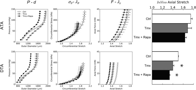
Passive biaxial mechanical behaviors from ascending (ATA – top row) and descending (DTA – bottom row) thoracic aortas: mean pressure–diameter data (first column) and associated circumferential Cauchy stress–stretch data (second column) for cyclic P-d tests plus mean axial force–length data for cyclic f-l tests performed at 100 mmHg (third column) and individual values of the in vivo axial stretch (bar plot – fourth column). Data are shown for three groups: controls (Ctrl), tamoxifen-induced disruption of Tgfbr2 without dissection (Tmx), and tamoxifen-induced disruption with daily treatment with rapamycin (Tmx+Rapa). Disrupted TGF-β signaling caused differential effects in the two aortic locations: loss of distensibility (with normal extensibility) in ATAs and loss of extensibility (with normal distensibility) in DTAs. Rapamycin neither prevented nor improved the passive biomechanical changes, despite reducing the diameter with respect to Tmx. * indicates p < 0.05 with respect to Ctrl samples from similar aortic locations.