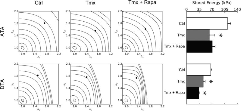
Figure 3.

Passive elastic energy storage during biaxial mechanical testing of ascending (ATA – top row) and descending (DTA – bottom row) thoracic aortas for three groups: Ctrl (first column), Tmx (second column), and Tmx+Rapa (third column). Iso-energetic contour plots highlight biaxial strain dependencies whereas individual values reveal in vivo states (bar plot – fourth column). Each iso-energy level is indicated as a curve (with values of 0.1, 1, 5, 10, 20, 40, 60, 100, 250, and 500 kPa), while in vivo values are shown as filled circles. ATAs normally store more energy than DTAs due to a higher elastin content and nearly equi-biaxial loading. Loss of TGF-β signaling decreased energy storage in both regions and rapamycin neither prevented nor restored this function (in ATAs, Tmx+Rapa energy storage is lower than Ctrl at p < 0.1 while being not significantly different from Tmx). * indicates p < 0.05 with respect to Ctrl samples from similar aortic locations.
