Figure 4.

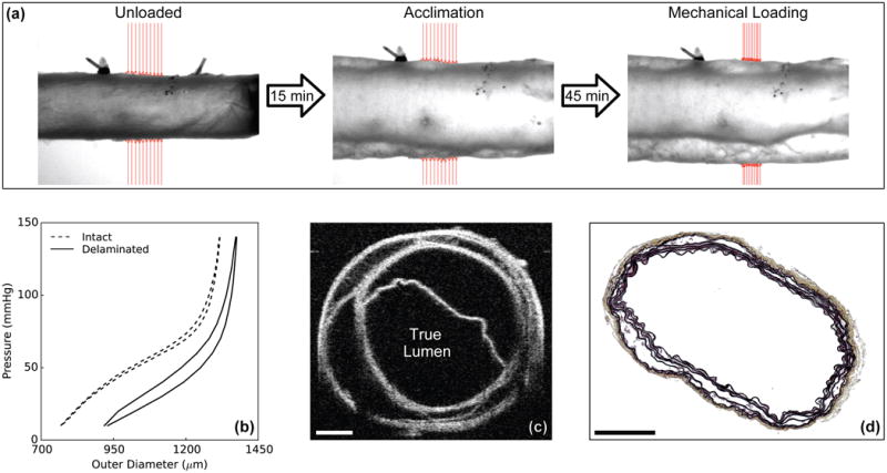
Illustrative data for mechanical testing under passive conditions of a rapamycin-treated, Tgfbr2 disrupted DTA (Tmx+Rapa). Video-images (a) reveal the initiation of an intramural delamination (separation of wall layers) upon static pressurization (acclimation), with progression during cyclic pressurization (mechanical loading from 10 to 140 mmHg repeated four times) despite the grossly normal appearance prior to loading (unloaded). Shown, too, are associated pressure-diameter data (b), an optical coherence tomographic image collected during testing (c), and a histological image obtained after testing (d). The cross-sectional images reveal complex, extensive separation of medial elastic layers with an otherwise intact adventitia (which allowed pressurization during mechanical testing). Mechanical data showed that delamination was associated with circumferential dilation (higher outer diameter) and increased hysteresis (higher energy dissipation), both consistent with the accumulation of interstitial fluid following delamination. Scale bars represent 200 μm.
