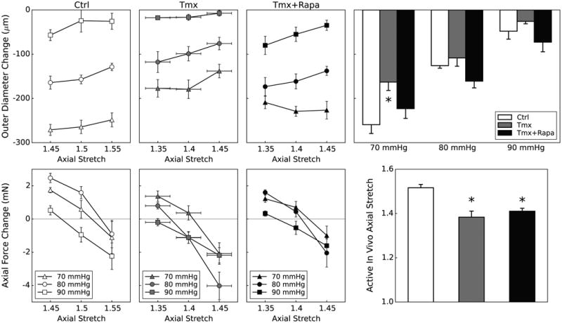
Figure 5.

Active biaxial mechanical data from descending thoracic aortas (DTA) contracted via 80 mM KCl for three groups: Ctrl (first column), Tmx (second column), and Tmx+Rapa (third column). Data in the first three columns show contraction-induced changes in outer diameter (top row) following 15 minutes of SMC activation and changes in axial force (bottom row), at three isobaric (70, 80, and 90 mmHg) and three axially isometric (around the vessel-specific in vivo stretch) conditions, thus yielding 9 different active states. The active in vivo axial stretch is calculated individually for each vessel at the three pressures as the value at which axial force remains constant upon contraction. Data in the far right columns summarize the primary biaxial findings: contraction-stimulated decreases in outer diameter (top) and values of active in vivo axial stretch (bottom). Passive and active in vivo axial stretches were indistinguishable (Table II) for each group. Tgfbr2 disruption diminishes SMC contraction at 70 mmHg, but rapamycin treatment tends to restore or augment this contractility at all pressures though without recovery of the preferred axial stretch. * indicates p < 0.05 with respect to Ctrl samples from similar aortic locations.
