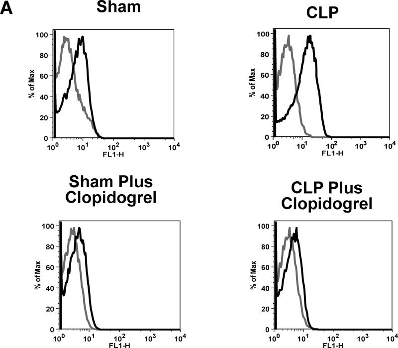
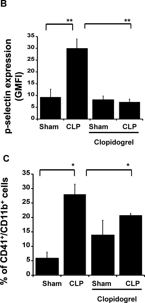
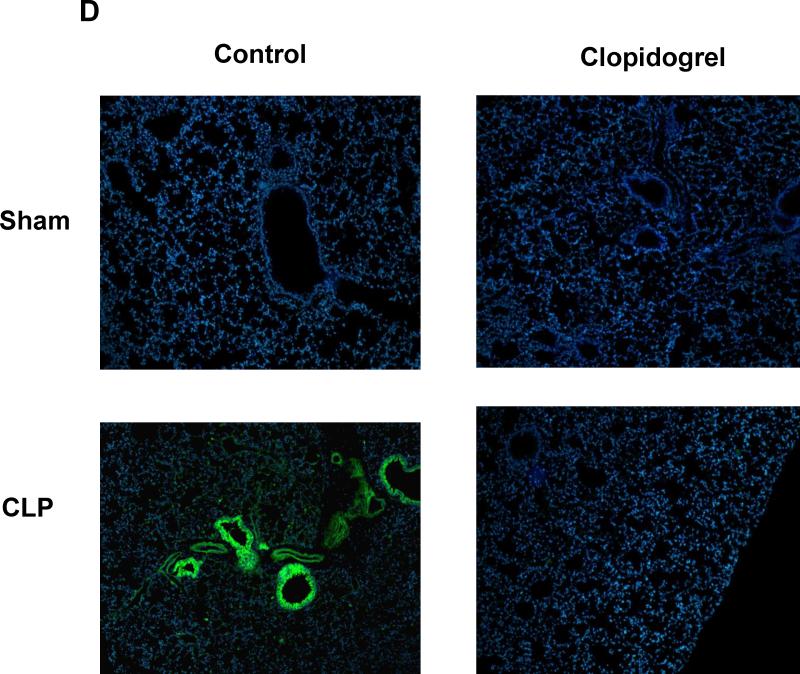
Figure 2. P-selectin expression and leukocyte-platelet aggregates were not elevated in clopidogrel treated mice during sepsis.
(A) and (B) Blood samples were collected by cardiac puncture in 3.8% sodium citrate (10:1), and P-selectin expression on platelet surface was analyzed through flow cytometry. Representative flow cytometry histograms are shown for CLP and sham controls in WT and KO animals. Isotype control is shown in gray and P-selectin stained samples in black. (C) Blood samples were labelled with antibodies against CD61 (platelet marker) and CD11b (leukocyte marker). Activated leukocytes were gated based on CD11b expression and cell shape, and data were analyzed as a percentage of aggregates expressing both CD41 and CD11b. Values are expressed as percentage of CD41+/CD11b+ cells, mean ± SEM (*p < 0.05; WT sham versus WT CLP and KO CLP versus WT, n = 6). (D) Representative images of CD41 staining (CD41: green; Nucleus: blue; 20x) for CLP and sham samples for both treated and untreated mice. Images are representative of 4 different experiments.


