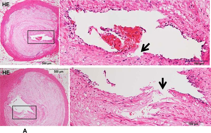
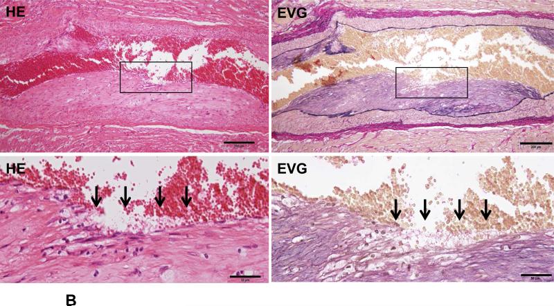
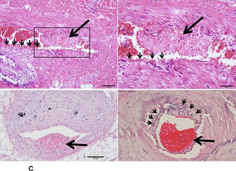
Figure 3. Coronary plaque rupture (A), erosion (B) and thrombosis (C) in Ang II-infused WHHL rabbits.
A. Two representative micrographs of coronary plaque rupture are shown. The lesions show marked lumen stenosis (>80%). At high magnification, the top lesion shows distinct disruption (arrowhead) associated with a thrombus. The bottom lesion shows an intimal split (arrowhead) in which some red blood cells are contained.
B. The surface of a fibrotic plaque was partially eroded or defective (arrows), covered by blood cells. The left panel shows HE staining and the right panel shows EVG staining.
C. Typical mural thrombi are shown in three coronary lesions. The coronary lumen was partially filled with a thrombus (arrows) which contains fibrin and blood cells (top and bottom panels). Please note that opposite to the thrombus, the lesion surface is eroded (arrowheads) and calcification is seen at the bottom of the lesions (top panels). Foam cells are accumulated beneath the thin cap of a vulnerable plaque (arrowheads) (bottom right).


