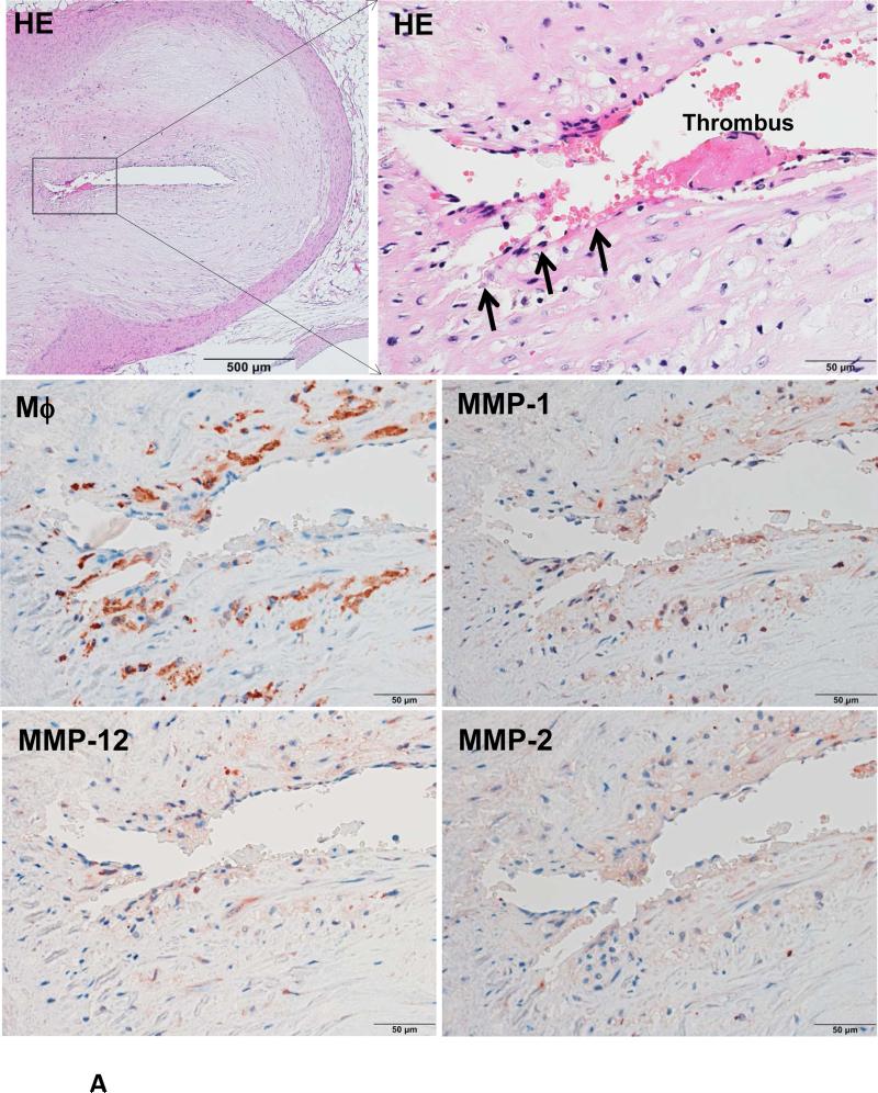
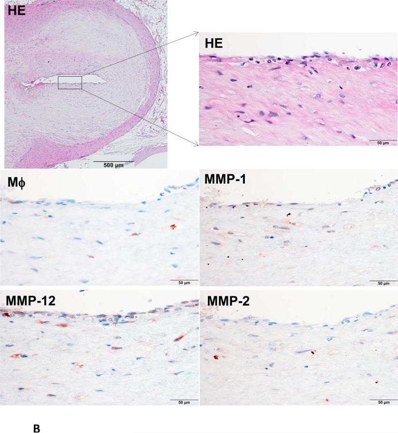
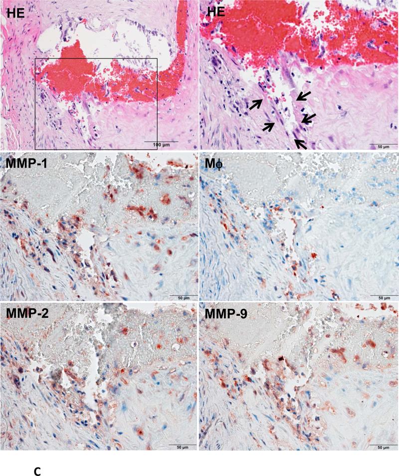
Figure 4. Immunohistochemical staining of macrophages and MMPs.
A representative eroded lesion (arrowheads) with a thrombus on the surface shows accumulation of a few macrophages and stained with MMP-1, -2 and -12 Abs (A). In the normal area adjacent to the erosion of the same artery, there are only a few macrophages that are stained with MMP Abs (B). Another representative ruptured lesion (arrowheads) associated with an occlusive thrombus shows accumulated macrophages with MMPs staining at the rupture area (C).


