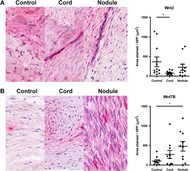
Fig. 3.
Representative pictures and quantification of staining of selected Wnt-proteins in Dupuytren’s disease tissue (cords, nodule) as compared to control tissue (unaffected transverse ligaments of the palmar aponeurosis). a Wnt2. b Wnt7b. * P < 0.05 by Kruskal-Wallis test, followed by post-hoc Dunn’s Multiple Comparisons test

