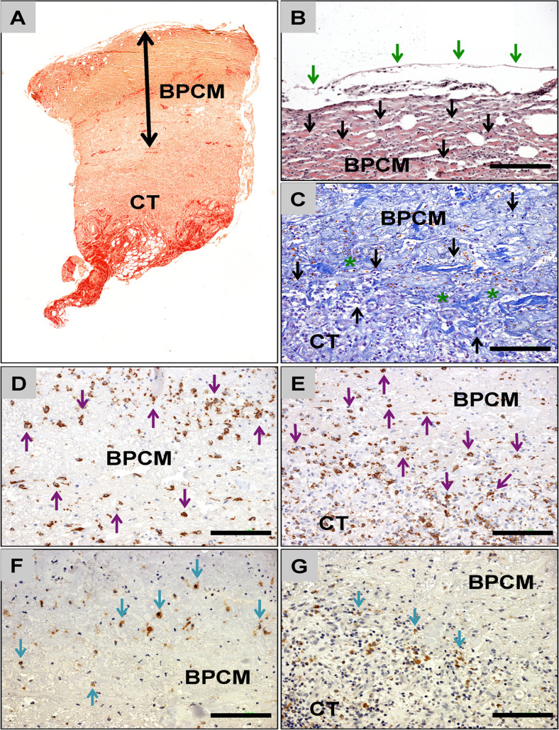
Fig. 8.
Exemparily histology of BPCM-based tissue regeneration of a medium-sized skin defect. a Overview of the implant area of BPCM (double arrows) implanted above the connective tissue (CT) of the wound base (Sirius-staining, „total scan“100× magnification). b The compact layer of the BPCM was covered by a thin and continuous layer of epidermal cells (green arrows). Mononuclear cells (black arrows) have invaded this material layer at this time point (HE-staining, 200× magnification, scale bar =100 μm). c The material of the spongy layer of the CM adjacent to the connective tissue of the wound ground (CT) was integrated into a connective tissue (asterisks) containing high numbers of mononuclear cells (black arrows) (Azan-staining, 200× magnification, scale bar =100 μm). d - g The CD-68 detection showed that most of the cells that invaded the compact d and the spongy e layer of the BPCM were macrophages (purple arrows). Only a few of the cells that have invaded the compact f and the spongy g components of the CM showed signs of TRAP-expression (d and e: CD68 immunohistochemistry, f and g: TRAP immunohistochemical staining, 200× magnification, scale bars =100 μm)

