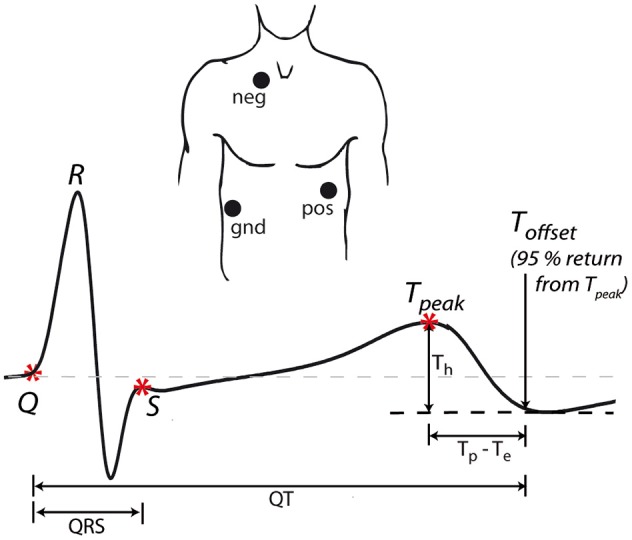
Figure 1.

ECG acquisition. Three electrodes (positive, negative, and ground) were placed in modified CS5 lead positions to obtain a derived Lead II. The example waveform shows ECG landmarks (*) and the measured intervals (QRS, QT, Tp–Te) and amplitudes (Th). For all intervals, the time of 95% return from T-peak to the minimum of the T wave was used as the end of repolarization.
