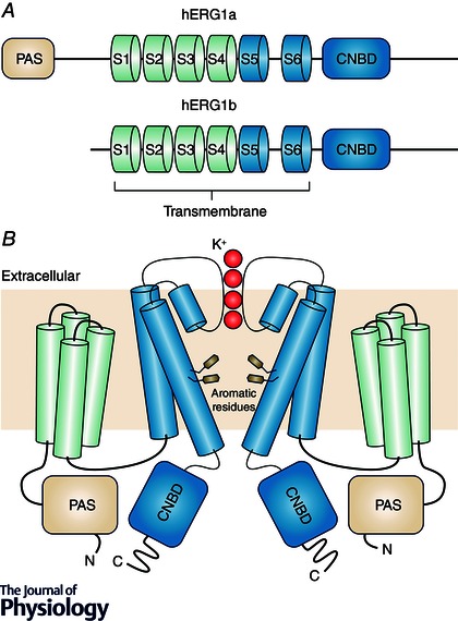
Figure 1. hERG channel structure .

A, domain structure of hERG1a (top) and hERG1b (bottom). hERG1a contains a cytosolic Per‐Arnt‐Sim (PAS) and cyclic nucleotide binding domain (CNBD), as well as 6 transmembrane helices S1–S6. The hERG1b N‐terminus lacks a PAS domain and instead contains a unique 35‐amino‐acid sequence. Domains are not drawn to scale. B, putative topology of the hERG K+ channel tetramer. For clarity, only one pair of opposing subunits comprising the hERG tetramer are shown. K+ ions passing through the channel selectivity filter are shown in red. Also shown are aromatic amino acids Y652 and F656 involved in drug‐induced hERG block.
