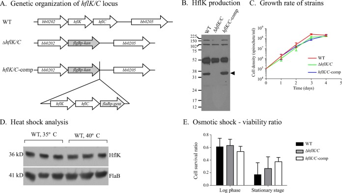
FIG 1 .
Phenotypic characterization of the ΔhflK/C mutant. (A) Genetic organization of the WT, mutant, and complemented strains at the hflK and hflC loci. (B) Immunoblot analysis of HflK synthesis by different B. burgdorferi strains. Antiserum raised against B. burgdorferi HflK reacts with a protein of the appropriate size (36 kDa [arrowhead]) from whole-cell lysates of WT and complemented strains, but not with a lysate of the ΔhflK/C mutant. (C) In vitro growth rate of the ΔhflK/C mutant compared to the WT and hflK/C-comp strains. The mean and standard deviation (SD) are displayed. (D) Immunoblot analysis of cell lysates from WT B. burgdorferi exposed to a 40°C heat shock treatment for 1 h or maintained at the standard growth temperature (35°C). FlaB levels are shown to demonstrate equivalent protein loads. (E) Cell viability of B. burgdorferi strains subjected to 1 N NaCl osmotic stress compared to untreated spirochetes. After treatment, cells were plated in solid BSK medium, and the viability ratio was determined by counting CFU of treated compared to untreated spirochetes. SD bars are shown; no significant difference in cell viability was detected among B. burgdorferi strains at either the log phase (one-way analysis of variance [ANOVA], P = 0.2850) or stationary phase (one-way ANOVA, P = 0.1478).

