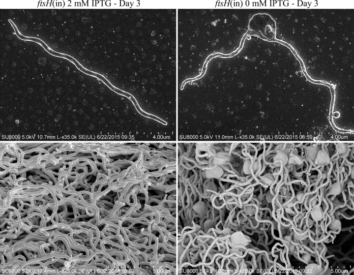
FIG 4 .
Morphology of FtsH-depleted B. burgdorferi cells compared to FtsH+ cells. Shown are representative scanning electron micrographs of the B. burgdorferi ftsH(in) strain grown for 3 days either in the presence of 2 mM IPTG and producing FtsH (left panel) or in the absence of IPTG, resulting in FtsH-depleted cells (right panel). Large membrane blebs are evident in FtsH-depleted cells.

